Radiology Referral Pathways Australia Guide
Navigate radiology referral pathways in Australia with criteria, urgency categories, state variations and decision nodes for clinicians.
You’re a GP (general practitioner) with a patient who needs imaging. The symptoms sound concerning, but the patient is clinically stable. The nearest imaging service uses one referral form, the hospital network uses another, and the booking team wants more detail before it can decide whether the request is routine or urgent. Meanwhile, the patient wants to know whether a scan can be arranged without waiting for a standard appointment.
Radiology referral pathways in Australia become practical rather than theoretical. The correct route depends on the clinical question, the requested modality, the urgency, the quality of the request, the Medicare rules that apply, and the requirements of the state, territory, hospital network or private provider receiving it. There isn’t one national pathway that overrides all of those variables.
Table of Contents
- How Radiology Referral Pathways Work in Australia
- What Makes a Valid Imaging Request in Australia
- Urgency Categories and Triage Decision Points
- Evidence Based Guidance Behind Appropriate Imaging Selection
- Modality Pathways and Common Clinical Scenarios
- State and Hospital Network Variations You Must Check
- From Request to Report and Image Access
- Access Challenges and Alternative Routing Options
- Quick Reference Matrix and Cross References for Clinicians
How Radiology Referral Pathways Work in Australia
A useful way to understand radiology referral pathways is to treat them as a series of decision nodes rather than a single line from GP to scan. At each node, someone decides whether the request is valid, clinically appropriate, urgent enough for escalation, suitable for the chosen modality, and compatible with the receiving service’s local process.
That matters because imaging sits inside a referral system that runs at national scale, and a request that stalls at any node is a patient waiting. Referral quality is a system issue, not a minor administrative detail.
Practical rule: A referral isn’t complete when it leaves the consulting room. It’s complete when the receiving service can identify the patient, understand the clinical question, assess urgency, select or confirm the modality, and return the result safely.
This guide is designed for clinicians, trainees and administrators who need a quick reference during a consultation or shift. Use the sections on request validity, urgency and modality selection for the clinical decision. Use the state and network section when the same request may be handled differently by a public hospital, private imaging provider or local health service.
The wider Australian medical education environment also depends on understanding local systems, because Australian medical training pathways expose doctors to different hospital workflows, referral conventions and assessment expectations. In practice, radiology referral pathways are clinically governed systems. They connect patient safety, resource use, triage, communication and funding.
What Makes a Valid Imaging Request in Australia
A patient arrives for imaging, but the request shows only a name and “scan.” The booking team cannot confirm the examination, the radiologist cannot judge the clinical question, and the service may need to seek clarification before triage or billing. A valid request prevents those separate problems from becoming one delay.
The six elements that prevent avoidable delay
Australian guidance describes a diagnostic imaging request as written, dated, legible and sufficiently specific. It should identify the patient, name the requesting clinician and provider number, and give a clear clinical justification for each examination. Queensland Health guidance on procedure requests also advises avoiding abbreviations that could be misread.
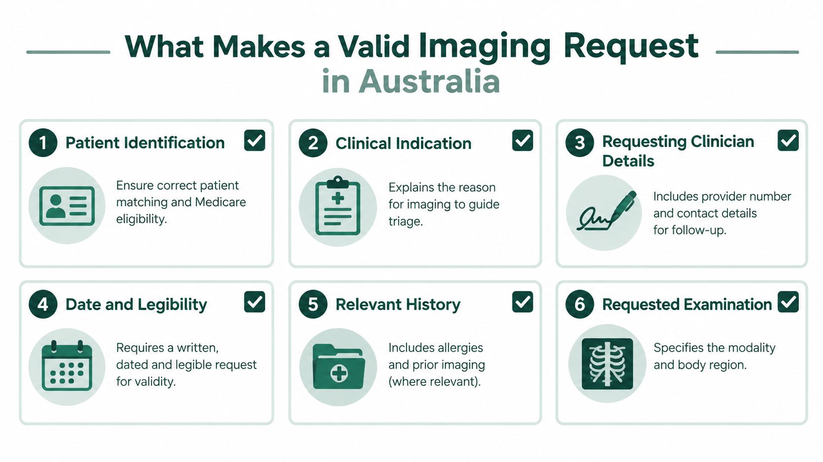
Use the request to give the receiving service a usable clinical map:
- Patient identification: Include identifying and contact details so staff can match the request, arrange communication and avoid mix-ups.
- Clinical indication: State the symptom, suspected diagnosis, relevant examination findings and the question the imaging should answer.
- Requesting clinician details: Provide the clinician’s name, provider number, contact details and required authorisation.
- Date and legibility: Make the request readable, signed where required and clearly dated. Missing or unclear details can affect booking and billing.
- Relevant history: Add information that may change triage or preparation, such as prior imaging, pregnancy status where relevant, allergies, renal considerations or previous surgery.
- Requested examination: Specify the modality and anatomical region. If the final examination may depend on radiologist review, explain the clinical reason rather than listing an unexplained scan.
These elements work together. Patient details identify the person, the clinical indication explains the problem, and the examination request gives the service a proposed route. History and contact details then allow the provider to modify preparation, clarify uncertainty or escalate the request when needed.
Why completeness affects Medicare eligibility
The request also supports Medicare compliance. For R-type imaging, Medicare benefits aren’t payable unless a requesting practitioner makes the request before the service begins, as described in the Medicare Online and Eclipse data elements guidance. A provider may therefore need to correct or confirm a deficient request before performing the service or claiming a benefit. Local public hospitals and private providers can apply further workflow requirements, even when the Medicare rule is satisfied.
Compare these examples:
- “CT abdomen, pain.”
- “CT abdomen and pelvis requested for acute right lower quadrant pain, fever and localised tenderness. Assess for suspected appendicitis. Include relevant prior abdominal surgery, pregnancy status where applicable, and the requestor’s provider details.”
The second request presents a clinical question and enough context for urgency review. It does not prove that CT is the best examination, but it makes the reasoning visible, allowing the receiving service to assess the request rather than guess.
Before sending: Read the request as if you were the booking clerk and radiologist. Without your consultation note, could they identify the patient, understand the concern and contact you?
Urgency Categories and Triage Decision Points
Urgency is not a decorative label. It tells the receiving service how quickly the request should be assessed and whether outpatient booking is an appropriate route at all. Local services may use different names or time bands, so the categories below are a clinical framework, not a substitute for the receiving network’s rules.
Compare the four common categories
| Category | Typical clinical position | Referral action |
|---|---|---|
| Emergency | A potentially life-threatening or time-critical problem | Use an emergency department or hospital pathway, and contact the relevant radiology team when immediate imaging is needed |
| Urgent | An acute but stable problem where delay could affect management | Mark the clinical reason clearly, provide contact details and confirm the provider’s booking process |
| Semi-urgent | A subacute problem requiring timely assessment but without immediate danger | Describe progression, examination findings and the reason routine booking is unsuitable |
| Routine | A non-urgent question where the result won’t change immediate management | Provide a complete request and follow the local outpatient booking process |
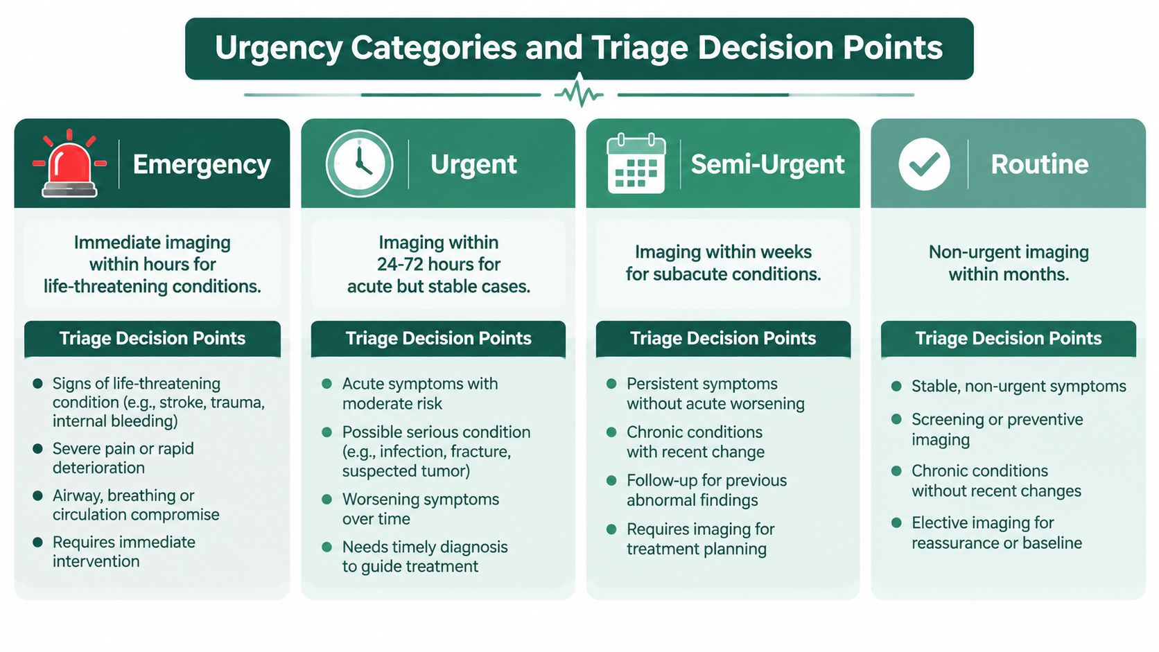
The first decision node is clinical stability. A patient with neurological deficit, haemodynamic instability, severe respiratory compromise or another time-critical presentation generally shouldn’t be placed into a routine outpatient pathway just because the imaging request is convenient to write. The appropriate route may be the emergency department, inpatient team or direct discussion with a radiologist or registrar.
The second node is whether the request explains the escalation. “Urgent” without supporting detail forces the service to infer the risk. Include the onset, progression, red flags, examination findings, suspected diagnosis and the management decision that depends on imaging.
When a phone call changes the pathway
Phone the radiology registrar or service when the diagnosis is time-sensitive, the patient may need a different modality, contrast or special preparation, or the request doesn’t fit a standard outpatient workflow. A call is also appropriate when the patient’s condition has changed after the request was sent.
The same symptom can move between categories. Stable, mechanical back pain with no concerning findings may follow a routine pathway. New weakness, saddle sensory change or bladder dysfunction requires immediate clinical assessment and a different escalation pathway. The imaging label alone doesn’t determine urgency. The combination of risk, stability, examination and management consequence does.
Incomplete context can also lower priority because the service can’t safely distinguish a time-critical presentation from a routine one. If the request is returned, treat that as a signal to clarify the clinical question, not as a booking inconvenience.
Evidence Based Guidance Behind Appropriate Imaging Selection
A request can be complete enough for processing yet still choose the wrong examination. Administrative completeness answers whether the service can process the referral. Clinical appropriateness asks whether the requested examination is the best way to answer the patient’s question.
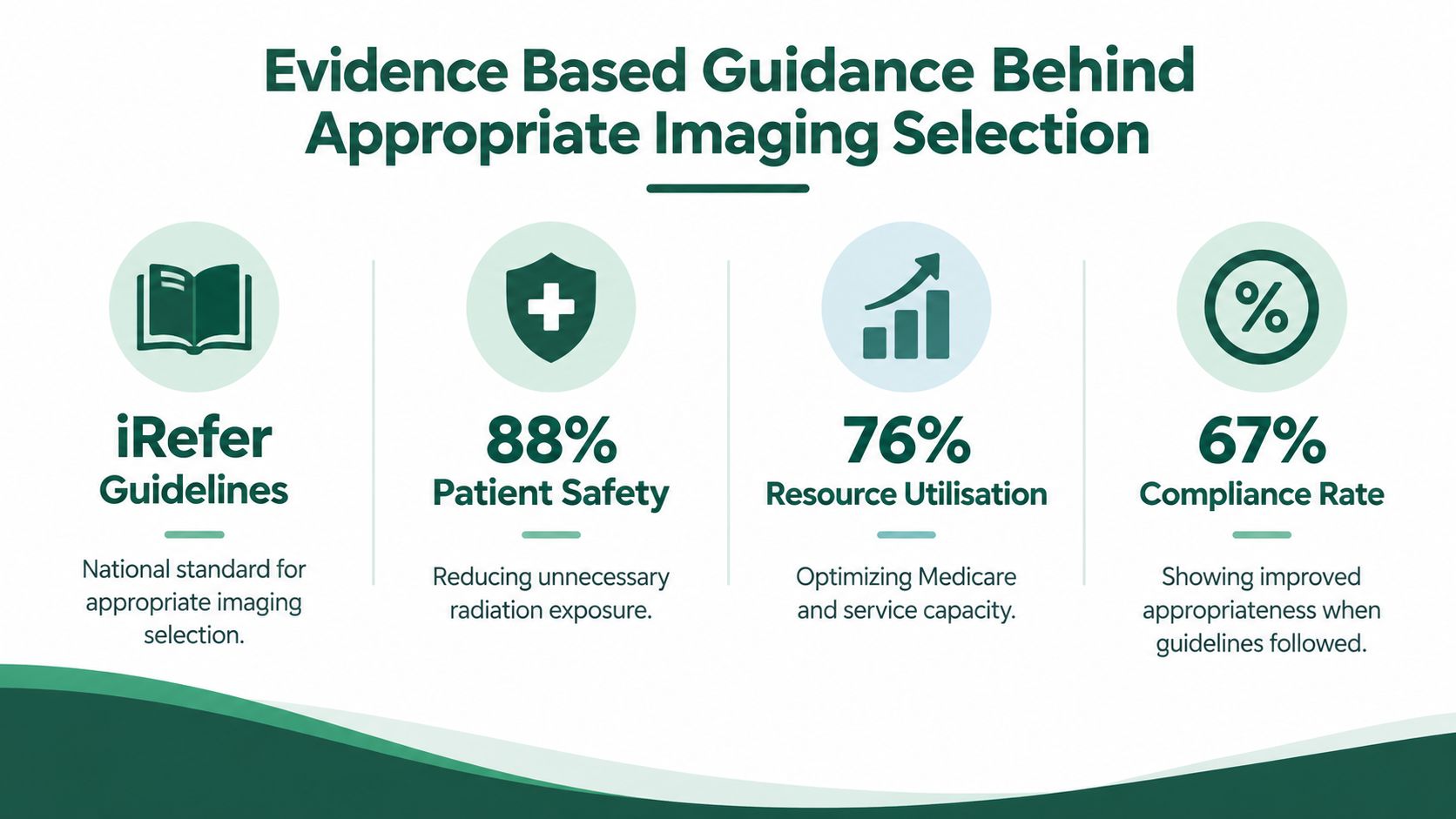
Australia and New Zealand use iRefer guidelines under an agreement between RANZCR (the Royal Australian and New Zealand College of Radiologists) and the RCR (the United Kingdom’s Royal College of Radiologists). RANZCR holds the exclusive licence for iRefer in Australia and New Zealand, and the RCR has published the guidelines since 1989 — see RANZCR’s own iRefer information. The guidance supports decisions about whether imaging is indicated and which modality is most suitable. It informs the referral decision, while state pathways and hospital network protocols determine how that decision is applied locally.
What the audit data teaches
A pilot audit of emergency-department X-ray referrals at a regional Queensland hospital found that only 24.7% complied with the Western Australian government’s diagnostic imaging pathways, the benchmark the auditors used, leaving 75.3% classified as unjustified. When the clinical details in the patient’s record were read alongside the referral, the unjustified rate fell to 49.2% — see Rawle and Pighills, Journal of Medical Radiation Sciences, 2018. Treat the size of that gap carefully rather than as a national rate: this was a pilot study covering an 11-day period at a single regional hospital.
The practical lesson is about documentation, not individual scorekeeping. A request may appear inappropriate when it leaves out examination findings or relevant history that supported the decision. Adding that context gives the reviewing radiologist a clearer basis to assess the indication, select an examination and assign an appropriate pathway.
Use guidance at the referral node
Start with the clinical question, rather than the scan a patient has requested. Ask:
- What diagnosis am I considering?
- Will imaging change immediate management?
- Is there a recommended first-line modality?
- What findings would make another test more useful?
- Have I documented the features supporting this choice?
For example, “headache” does not explain why neuroimaging is needed. Sudden onset, neurological findings, trauma, systemic symptoms or another defined concern gives the radiologist a basis for modality review and triage. A low-value request without a clinical question can cause delay, unnecessary radiation exposure or an examination that cannot answer the concern.
Guidelines support clinical judgement. They do not replace patient assessment, discussion with radiology or the local rules that apply to complex cases.
Modality Pathways and Common Clinical Scenarios
The modality should follow the clinical question, not the other way around. Each examination has strengths, limitations, preparation requirements and safety considerations, so the referral needs to give the radiology team enough information to confirm the route.
Plain radiography
Plain X-ray is often useful for an initial assessment of bones, joints and the chest. State the body region, side, mechanism or relevant history, and the suspected condition. “Chest X-ray” is less useful than a request that explains whether the concern is infection, heart failure, trauma or another clinical question.
Radiography uses ionising radiation, so pregnancy status and previous relevant imaging may matter. It may also be unsuitable as the only test when the suspected abnormality is poorly visualised on X-ray or when cross-sectional imaging is needed.
Ultrasound
Ultrasound is useful for many soft-tissue, vascular, pelvic and pregnancy-related questions and doesn’t use ionising radiation. The request should identify the region, symptoms, duration, examination findings and any relevant surgical or obstetric history.
A common error is requesting “abdominal ultrasound” without specifying the suspected problem. Right upper quadrant pain, jaundice, a palpable mass and urinary symptoms can lead to different protocols and preparation instructions.
CT
CT provides rapid cross-sectional imaging and is often important in acute care. The request should state the suspected diagnosis, anatomical coverage and whether contrast may be relevant. Include allergies, renal considerations, pregnancy status where relevant, prior surgery and the patient’s clinical stability.
For suspected pulmonary embolism, the pathway depends on the overall clinical assessment and local protocol. A stable patient may need a structured outpatient or emergency assessment route, while respiratory compromise, haemodynamic instability or other red flags should direct the patient to urgent hospital care rather than routine booking.
MRI
MRI gives detailed soft-tissue information without ionising radiation, but access, safety screening and preparation requirements vary. Tell the service about implanted devices, metal exposure, prior surgery, pregnancy where relevant and the exact clinical question.
The modality is not automatically the best choice for low back pain or headache. The presence of red flags, neurological findings, trauma, cancer history or systemic illness should determine escalation and the need for discussion.
Nuclear medicine
Nuclear medicine examines function using a tracer. The request should explain the clinical question, relevant medications or therapies, pregnancy or breastfeeding considerations where applicable, and any preparation requirements. A suspected functional disorder may follow a different route from a request for anatomical detail.
For every modality, the same decision applies: define the question, document the context, check safety, then confirm the local booking and triage route.
State and Hospital Network Variations You Must Check
Australian radiology referral pathways vary by state, territory, local health district, hospital network and provider. A request accepted by one service may be returned by another because the receiving organisation uses a different form, portal, triage rule, booking hub or image-access system.
Where the differences appear
| Area | What may vary locally |
|---|---|
| Referral form | Mandatory fields, accepted signatures, templates and supporting documents |
| Electronic submission | Health service portal, secure messaging platform, e-referral process or fax fallback |
| Booking | Direct booking by the patient, centralised hospital hub or provider-managed scheduling |
| Triage | Local priority definitions, radiologist review and escalation requirements |
| Reports and images | Referrer portal, hospital record, secure message, disc, patient portal or linked viewer |
| Eligibility | Public hospital catchment, clinician type, Medicare requirements and provider-specific rules |
These variations aren’t evidence that one network is careless and another is correct. They reflect different services, clinical populations, governance arrangements and information systems. A public outpatient department may require a local form and catchment details, while a private provider may accept a referral through a secure electronic channel but still apply its own modality and Medicare checks.
A reliable local lookup method
Before sending the request, check the receiving service’s own instructions. Start with the relevant state or territory health service, local health district imaging page, hospital intranet or approved HealthPathways entry. If the patient is being referred privately, check that provider’s current requirements rather than relying on a form saved from a previous referral.
The Australian medical exam and training landscape also demonstrates why local terminology matters for clinicians moving between jurisdictions. The same habit applies clinically: name the exact network and follow its current workflow.
Localisation check: Confirm the receiving site, accepted submission method, referral form, booking contact, urgency terminology and method for accessing the final report and images.
Don’t average local differences into a fictional national process. If you’re uncertain, phone the imaging department before the patient leaves, particularly when the request is urgent, the modality is specialised or the patient may not meet the network’s outpatient criteria.
From Request to Report and Image Access
Once the request is sent, the pathway usually moves through several operational checkpoints. Understanding them helps explain why a clinically sensible referral can still be delayed.
The workflow after submission
First, the service receives the request through an approved channel. Booking staff or a clinical reviewer check the patient’s identity, requestor details, examination, indication and urgency. A radiologist may redirect the modality, ask for further information or recommend a different pathway.
The service then contacts the patient or referrer about preparation, consent, appointment details and safety screening. Preparation can affect whether the examination goes ahead, so instructions about fasting, medication, hydration or arrival requirements need to reach the patient clearly.
After imaging, a radiologist interprets the study and issues a report. The report and images return through the provider’s agreed system, which may be a secure referrer portal, hospital record, electronic message or another approved viewer. The treating clinician remains responsible for reviewing the result, acting on it and communicating clinically important findings to the patient.
Common failure points and practical fixes
- Missing provider number: Add the requestor’s provider details before submission, then check that the service can contact the clinician.
- Weak justification: State the symptoms, findings, suspected diagnosis and management question rather than using a broad symptom label.
- Wrong modality: Describe the clinical problem and invite radiology review where the best test isn’t certain.
- Unsafe transmission: Use the provider’s approved secure channel. Don’t send identifiable clinical information through an informal method.
- Unclear result destination: Confirm who will receive the report, especially when the patient is seen across practices or hospital services.
The fastest workflow is usually the one that resolves uncertainty early. A complete request reduces clarification calls, but it doesn’t remove the need for clinical communication when urgency or modality choice is difficult.
Access Challenges and Alternative Routing Options
A patient may understand that imaging is needed but still struggle to obtain the request. GP availability can be limited, while Medicare-linked imaging generally requires a valid request from a medical practitioner. That creates a practical tension between access speed, clinical governance and benefit eligibility.
Some services market rapid telehealth e-referrals as a way to connect patients with a medical practitioner who can assess the problem and issue a request. That may improve access in selected circumstances, but a rapid electronic request still needs appropriate clinical assessment, sufficient documentation and compliance with the receiving imaging service’s requirements. Speed doesn’t make an incomplete or clinically unsuitable referral valid.
When alternative routing may help
Telehealth may be reasonable when the patient can be assessed safely through that format, the presentation isn’t time-critical, and the practitioner can obtain the information needed for a sound imaging decision. It isn’t a substitute for emergency assessment when red flags or instability are present. Nor should a patient use telehealth to bypass a required hospital or specialist pathway.
Referral and rebate arrangements also change over time. Treat the current service instructions and the current Medicare Benefits Schedule as authoritative, and avoid promising a particular Medicare outcome before the provider confirms eligibility.
A more tiered system
The old assumption that every pathway runs directly from GP to scan is becoming less reliable. Some providers have introduced specialist-review fees for referrals from allied specialists, indicating that referral acceptance and clinical review may depend on who initiated the request and which service receives it.
For clinicians, the decision is straightforward even when the system isn’t:
- Emergency presentation: Use emergency or hospital escalation, not a consumer-facing rapid referral route.
- Stable patient needing diagnostic clarification: Consider an appropriate medical consultation, including telehealth where clinically suitable.
- Specialist or allied referral: Check whether the imaging provider requires a medical practitioner’s request or applies a review process.
- Medicare-sensitive service: Confirm the request was made before imaging and contains the required details.
Alternative routing should improve access without weakening clinical responsibility.
Quick Reference Matrix and Cross References for Clinicians
Use the matrix as a prompt, not as a replacement for assessment or local instructions. The preferred pathway can change when examination findings, pregnancy, prior surgery, implanted devices, renal concerns or clinical deterioration alter the risk.
Radiology Referral Quick Lookup Matrix
| Clinical Indication | Preferred Pathway and Urgency Cue | Must Include in Request | Cross Reference |
|---|---|---|---|
| Suspected fracture or acute chest concern | Plain radiography may be an initial route. Escalate when instability, major trauma or significant respiratory compromise is present | Body region, side, mechanism, onset, examination findings and relevant pregnancy information | Plain radiography and urgency |
| Right upper quadrant pain or jaundice | Ultrasound may be appropriate, with urgency based on systemic illness and clinical stability | Pain location, duration, fever, jaundice, examination findings and relevant surgical history | Ultrasound and local booking rules |
| Suspected pulmonary embolism | Follow the local acute-care protocol. Don’t send an unstable patient through routine outpatient booking | Symptoms, observations, risk assessment, examination findings and the reason for the requested pathway | CT, contrast discussion and urgency |
| Low back pain | Imaging depends on red flags, neurological findings and suspected pathology. Routine pathways may suit uncomplicated presentations | Duration, neurological examination, red flags, trauma, cancer or systemic features where relevant | MRI and evidence-based selection |
| Headache | Urgency and modality depend on onset, neurological findings, trauma and systemic features | Onset pattern, examination, neurological findings, associated symptoms and suspected diagnosis | CT or MRI and triage |
| Functional or tracer-based question | Nuclear medicine pathway with preparation and safety screening | Clinical question, relevant treatment, pregnancy or breastfeeding considerations and preparation details | Nuclear medicine and provider instructions |
The final pre-send check
Confirm the patient’s identity, the requested examination, clinical justification, date, legibility, requestor details, provider number and contact pathway. Then ask whether the urgency label matches the patient’s actual risk and whether the chosen service is the correct local destination.
When a case is difficult, phone the radiologist with a concise summary: patient stability, key findings, suspected diagnosis, relevant contraindications, what has already been done and the management decision that depends on imaging. That conversation often resolves uncertainty more safely than sending a vague request and waiting for it to be returned.
For clinicians building broader knowledge across Australia’s health system, Australian medical specialties provide useful context for how specialty roles and local pathways intersect. Keep the matrix available, but always verify the receiving network’s current requirements before submission.
CliniRoo is building question banks, interview stations and format guidance mapped to the Australian pathway, for medical students, junior doctors and college trainees. Nothing is on sale yet. If you want to be told when there is something to use, CliniRoo takes an email address and does nothing else with it.